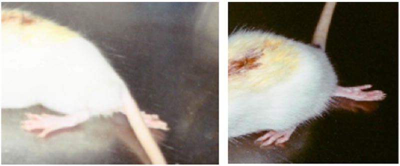
Figure 4.

Two representative sub-hemisectioned animals show impairment of their right hindlimb. One day post spinal cord microsurgical sub-hemisection show animals with sensory-motor impairment of their right hindlimbs, inducing the plantar side of the foot to face up and the contralateral hindlimb to the lesion to appear almost as in non-lesioned animals. The contralateral hindlimb shows a little sensory deficit, but the plantar side of the foot is facing down.
