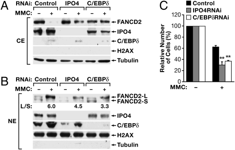
Fig. 6.
IPO4 augments nuclear localization of FANCD2 and cell survival in response to MMC. MDA-MB-468 cells were transiently transfected with siRNA against IPO4 or C/EBPδ or with scrambled oligos as control. MMC (500 ng/mL) was added 24 h later, and cells were incubated for another 20 h before preparation of (A) cytoplasmic (CE) or (B) nuclear (NE) cell extracts, followed by Western blot analysis of protein expression as indicated. The purity of cytoplasmic versus nuclear fractions also is shown in Fig. S7. (C) MDA-MB-468 cells were transfected as above and were placed in 96-well dishes at 5,000 cells/well 8 h later. The next day, MMC (500 ng/mL) was added, and cell viability was assessed 48 h later. Data are mean ± SEM of three experiments, each performed in triplicate, and relative to cells before treatment (set at 100%). **P < 0.01 relative to control siRNA.

