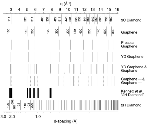
Fig. 3.
Electron diffraction peaks calculated for 3C cubic diamond, 2H hexagonal diamond, graphene, and graphene/graphane mixture compared to those measured from presolar graphene as well as graphene and graphene/graphane aggregates in carbon spherules isolated from Santa Rosa Island, CA. In the electron diffraction patterns identified as hexagonal diamond by Kennett et al. (16), asymmetrically doubled diffraction lines are clearly evident in their Fig. S2B as well as discernible in both their Fig. 2F and Fig. S2A. Peaks measured from the doubled diffraction lines in Fig. S2B of Kennett et al. (16) are shown (we calibrated the reported {100} reflection to 2.189 Å, and the line widths represent the error in our measurement). Kennett et al. (16) identified their Fig. S2C as hexagonal diamond; however, it is also consistent with diffraction normal to the graphite basal plane; graphite {100} (2.139 Å) is close to that of lonsdaleite {100} (2.182 Å).

