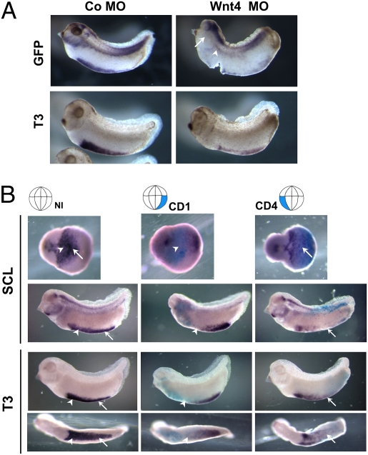
Fig. 3.
Depletion of Wnt4 affects formation of VBI. (A) Wnt-reporter embryos were injected at two-cell stage with 10 ng Co MO or Wnt4 MO in marginal zones, cultured to stage 32, and hybridized in situ with probes for dGFP and blood marker T3-globin (T3). Wnt4 morphant lost most of the GFP and T3-globin signals at the VBI. GFP transcripts were totally abolished at the DLP in Wnt4-depleted embryos (Right, arrowhead). Note that the GFP signal in the morphant embryo was also abrogated in the branchial arches (Upper Right, arrows) and lateral plate mesoderm area, which normally show abundant Wnt4 expression (Fig. 2G, arrow and arrowhead). (B) Targeted depletion of Wnt4 at CD1 or CD4 blastomeres (Upper, highlighted) by injection of Wnt4 morpholino ablates expression of hematopoietic cell markers SCL and T3-globin (violet blue) in corresponding VBI compartments. Arrowheads and arrows indicate aVBI and pVBI, respectively. Cytoplasmic β-gal RNA was coinjected as a lineage tracer (cyan blue).

