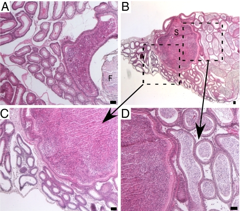
Fig. 4.
Spermatocele formation in mutant mice. The epididymides of adult Amhr2-Cre/+;Ctnnb1Δ(ex3)/+ mice were analyzed by H&E. Obstructive azoospermia were observed upstream of retained fibrotic (F) MD tissue (A). (B) An epididymis from another mutant with a large spermatocele (S). The boxes show the areas enlarged in C and D, which show the absence of sperm in the duct downstream of the spermatocele and the presence of sperm in the duct upstream, respectively. (Scale bar = 50 μm.)

