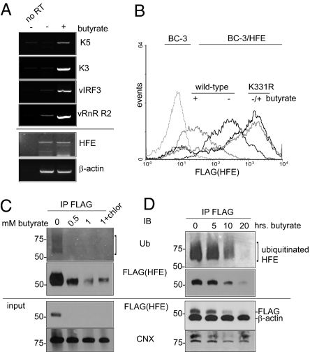
Fig. 5.
Lytic KSHV degrades HFE by targeting K331. (A) Primary effusion lymphoma BC-3 cells untreated or treated with sodium butyrate (1 mM) to induce lytic phase KSHV. After 24 h, total RNA was prepared. Transcript expression was analyzed by RT-PCR and visualized by agarose gel. A no reverse transcriptase enzyme control (no RT) was included to show that transcript detection was dependent on first-strand cDNA synthesis. (B) BC-3/HFE wild-type and K331R cells, left untreated (−) or treated (+) with butyrate (1 mM) for 24 h, were analyzed for binding of anti-FLAG antibody by flow cytometry. Dashed line, binding of FLAG antibody to parental BC-3 cells. (C) Detection of ubiquitinated HFE in KSHV positive cells. Shown are BC-3/FLAG-HFE wild-type cells left untreated, treated with butyrate (0.5 and 1 mM), or treated with butyrate and chloroquine (1 mM and 10 μM, respectively). After 24 h, cell lysates were subjected to anti-FLAG antibody IP, followed by IB with anti-Ub. IB using rabbit anti-FLAG showed that HFE was degraded efficiently in butyrate-treated cells. IB for anti-CNX was used as a loading control. (D) BC-3/HFE wild-type cells treated with butyrate (1 mM) for 0, 5, 10, and 20 h, analyzed by anti-FLAG IP and anti-Ub IB as in C. IBs for β-actin and CNX were used as controls.

