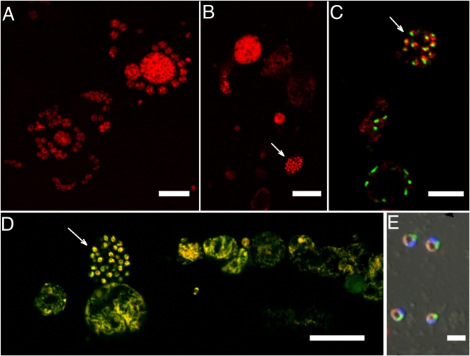
Fig. 3.
FISH of Nephromyces and Toxoplasma using apicomplexan-specific (Api-L) and Nephromyces-specific (Neph-1) SSU rRNA probes. Both probes were bound to fluor Cy3 (red). (Scale bars: A, B, D, 20 μm; C, 10 μm; E, 3 μm.) (A) Toxoplasma, Api-L; (B) Nephromyces from M. occidentalis, Api-L. Arrow indicates spores or flagellated cells. All other cells are uncleaved sporangia or trophic stages. (C) Nephromyces from M. manhattensis, Neph-1 (with eubacterial probe, EUB 338, linked to the fluor BODIPY FL (green). Toxoplasma did not bind to the Neph-1 probe. Arrow indicates spores or flagellated cells. All other cells are trophic stages. (D) Nephromyces from M. manhattensis, Api-L [merged with general eukaryote probe, EUK-516, linked to green (Fluos) fluor. Yellow represents binding to both Api-L and EUK-516]. Arrow indicates spores or flagellated cells. All other cells are uncleaved sporangia or trophic stages. (E) Nephromyces spores, Api-L, with EUB 338-BODIPY FL (green), and DAPI (blue).

