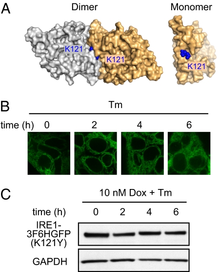
Fig. 3.
Disruption of IRE1’s ER–luminal dimerization interface blocks clustering and RNase activity of human IRE1. (A) Crystal structure of human IRE1 luminal domain dimer and monomer (PDB ID code 2HZ6). Structure identifies K121 as tightly packed residue at dimerizing interface. (B) IRE1-3F6HGFP(K121Y) mutant construct localization with Dox (10 nM) and Tm (5 μg/mL) treatment for indicated hours. (C) Level of IRE1-3F6HGFP(K121Y) protein expressed is examined via immunoblotting against total 3F6HGFP-IRE1 under 10 nM Dox induction.

