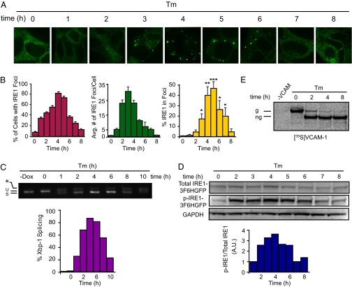
Fig. 4.
Sustained ER stress triggers disassembly of IRE1 clusters and IRE1 dephosphorylation. Stably transformed T-REx293 cells were grown in medium containing 10 nM Dox. At time 0, medium was replaced by medium containing no Dox but 5 μg/mL Tm to induce ER stress. (A) IRE1-3F6HGFP localization over 8-h time course. (B) Quantification of time course experiment. Percentage of cells with IRE1 foci, average number of IRE1 foci per cell, and percentage of IRE1 in foci were determined as described in Methods. Error bars represent SEM (*P < 0.05, **P < 0.005, ***P < 0.0005). Statistical significance of the difference between later time points and 0 h is indicated. (C) Xbp-1 mRNA splicing was determined by RT-PCR and quantified. (D) Total and phospho-IRE1-3F6HGFP protein levels were determined by immunoblotting. Histograms represent ratio of phosphoylated-IRE1-3F6HGFP and total IRE1-3F6HGFP. (E) T-REx293 cells were pulse-labeled for 1 h with [35S]methionine at the indicated times after beginning of the Tm treatment. Radiolabeled VCAM-1 (g, glycosylated; ng, nonglycosylated) was detected after immunoprecipitation and SDS gel electrophoresis.

