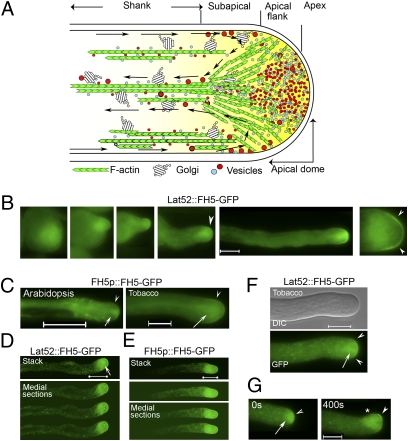
Fig. 1.
FH5-GFP is localized to the pollen-tube tip. (A) A schematic representation of angiosperm pollen-tube actin cytoskeleton and the apical inverted cone region (or “clear zone”) of vesicles. Arrows trace the “reverse-fountain” cytoplasmic streaming pattern. (B–F) Localization of formin homology-5 (FH5) promoter (FH5p) and the pollen-specific Lat52 promoter-expressed FH5-GFP to the apical and subapical membrane of Arabidopsis and tobacco elongating pollen tubes. (B) Emerging to elongating (Left to Right) Arabidopsis tubes. (D and E) Whole-tube projection of Arabidopsis tubes (Upper) by confocal imaging; three medial sections are shown (with increased γ to reveal signal from the shank, where no cell membrane-associated GFP-signal was detected). (F) Typical FH5-GFP localization pattern of prominent association with the apical and subapical, tip-focused collection of vesicles, and vesicular congregates that streamed along the cytoplasm (Movie S1). (G) A reorienting tobacco tube. Arrowheads highlight the apical flank and subapical regions; arrows indicate inverted cone region; asterisk in G indicates where the tip was at 0 s. See Fig. S2 for additional details and FH5p:GUS expression in transformed Arabidopsis pollen. Arabidopsis tubes were from stably transformed pollen. Tobacco tubes were transiently transformed by either 2 μg of Lat52::FH5-GFP or 10 μg of FH5p::FH5-GFP; elongating pollen tubes showed comparable FH5-GFP signals. (Scale bars, 10 μm.)

