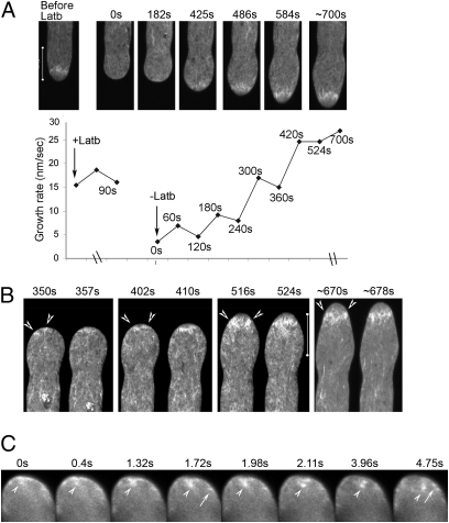
Fig. 4.
FH5 nucleates assembly of the subapical actin structure. Pollen tubes shown were transiently cotransformed by FH5 and actin reporter. (A and B) Subapical actin structure and growth recovery after Latb (12.5 nM) treatment. (A) Selected images from a confocal time series (Upper) and growth rates (Lower). (B) Subapical emergence of actin filaments (arrowheads), and their merging to form a structure spanning the central cytoplasm throughout the entire recovery period. More individual still frames and the entire time series are shown in Fig. S5 and Movie S6. (C) Selected images from a wide-field time series (after deconvolution) from a tube observed (∼5 min after Latb washout) when apical actin assembly was recovering from the apical membrane (Movie S7). Arrows, emanating actin filaments; arrowheads, dislodging actin fragments.

