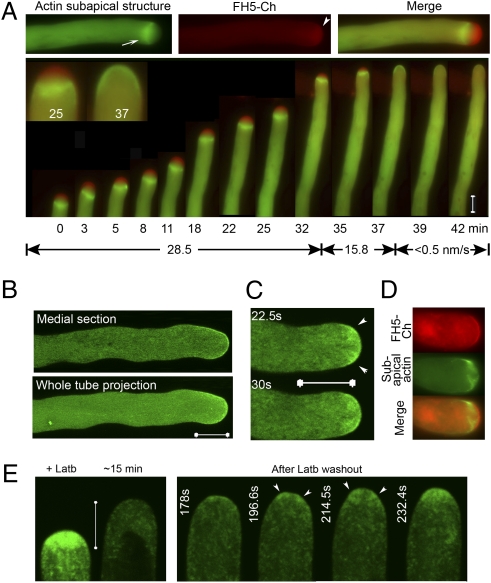
Fig. 5.
FH5-nucleated actin filaments support subapex-targeted vesicular trafficking. (A) Wide-field images monitoring growth of a tobacco pollen tube cotransformed by FH5-Ch and actin marker. (Upper) A representative image in individual channels and merged. Arrow, subapical actin structure; arrowhead, apical membrane and inverted cone region. The red signal was stretched equally in all merged images to illustrate the FH5-Ch labeled vesicular zone. Fig. S6 A and B show analysis details for the prominence of the subapical actin structure (SAC:Cyt) and of the apical vesicular zone (AV:Cyt) during transition from rapid to decelerated growth for this and other similarly transformed tubes. (B) A FH5-induced apical actin rim during the growth transition period. (C) A FH5-induced subapical vesicle targeting pattern (arrowheads) during the growth transition period. Two consecutive medial confocal sections are shown (7.5 s apart). (D) Subapex-focused actin filaments during a FH5-Ch induced growth transition. (E) Reaccumulation of vesicles during recovery from Latb-induced (+Latb) vesicle dispersal. Growth rate accelerated from < 7 nm/s to > 21 nm/s before and after 178 s. Arrowheads indicate subapical region. (Scale bars, 10 μm.)

