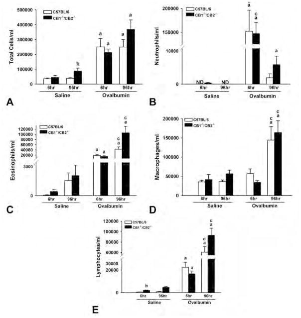
Figure 2.
Ovalbumin (OVA)-induced inflammatory cells in the bronchoalveolar lavage fluid. Lungs from saline- or OVA-treated wild-type and CB1/CB2 null mice were washed twice with saline, and differential staining of cell types was performed. Cells were enumerated using a 40× objective. (A) Total. (B) Neutrophils. (C) Eosinophils. (D) Macrophages. (E) Lymphocytes. (a) p < .05 versus respective saline. (b) p < .05 treatment-matched genotype difference. (c) p < .05 treatment-matched time difference.

