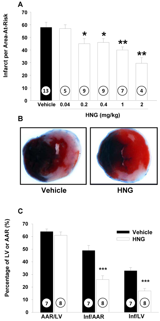
Figure 2.
HNG decreases myocardial infarct size in mice following MI-R: A) myocardial infarct size in mice receiving doses of HNG ranging from 0.2 to 2 mg/kg. HNG significantly decreased myocardial infarct size compared with vehicle in a dose dependent manner. B) Representative mid-ventricular photomicrographs of mouse hearts are shown after 45 min of myocardial ischemia and 24 hr of reperfusion. Areas of the myocardium that appear blue represent the areas of myocardium not at risk for infarction. In contrast, the areas of myocardium that stain red (i.e., TTC positive) represent viable myocardium that was at risk for infarction. Myocardium that appears pale (i.e., TTC negative) indicates areas of myocardium at risk that are necrotic (i.e., infarcted). C) HNG administered at the time of reperfusion decreased infarct size significantly compared to AAR. Values are means ± SE. Numbers inside bars indicate the number of animals investigated in each group. **p <0.01; ***p <0.001 vs. vehicle.

