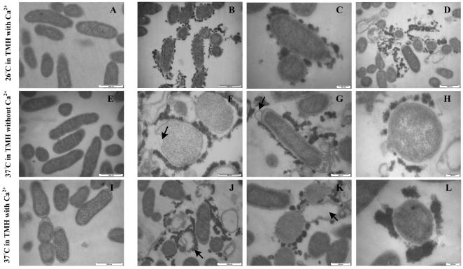
Figure 7. RovA mutation leads to alteration of the bacterial membrane.
Bacterial strains were either grown in TMH medium with 2.5 mM calcium at 26°C to the early stage of the stationary phase (A, wild type strain; B, C and D, ΔrovA mutant) or grown in the same medium to an OD600 of 0.3 and then transferred to 37°C for 3 h (E, wild type strain; F, G and H, ΔrovA mutant); or grown in TMH without calcium at 26°C to an OD600 of 0.3 and then transferred to 37°C for 3 h (I, wild type strain; J, K and L, ΔrovA mutant). Bacterial cells were harvested and subjected to transmission electron microscopy observation. Bacterial cells of the ΔrovA mutant were shown to be surrounded by electron dense particles in B, C, F, G, H, K and L. Arrows in J and K indicate the disrupted cells, and the covering of electron dense materials around the disrupted cells could be clearly observed. Arrows in F and G indicate bubbles on the bacterial membrane. Bars indicate 200 nm in C, H and L; 500 nm in F, G and K; 1,000 nm in A, B, D, E, I and J.

