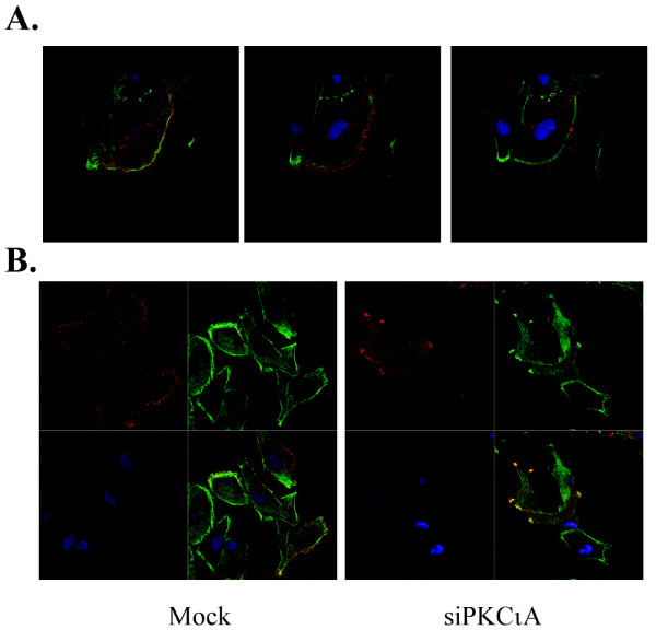
Figure 5.
Confocal microscopy of Lgl and non-muscle myosin IIA in PKCι-depleted cells. A. Immunocytochemistry for Flag-tagged Lgl (red) and non-muscle myosin IIA (green) was performed on U87MG cells transduced with Flag epitope-tagged Lgl cDNA. Nuclei were stained with DAPI (blue). Three serial confocal optical sections are shown for a cell with a distinct leading edge, with the section closest to the substratum on the left. B. Confocal images of U87MG cells transduced with Flag epitope-tagged Lgl cDNA that were either mock-transfected (left) or transfected with an RNA duplex targeting PKCι (right). Flag-Lgl (red), non-muscle myosin IIA (green) and DAPI (blue) images are shown separately and as a merged image in the bottom right quadrant.

