
FIGURE 1. TRHR1 receptors in the dorsal medulla.
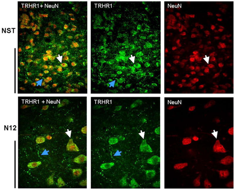
Example of double IHC staining for TRHR1 (green) and NeuN (red) in the NST (top row) and the hypoglossal nucleus (N12; bottom row) indicates that TRHR1-ir is superimposed on neurons (white arrows) and is also present on fibers and varicosities (blue arrows) in these areas. In both rows, NeuN IHC staining alone is seen in the far right; middle panel represents TRHR1 staining alone; and far left panels are merged images of the double stained sections. A similar distribution of TRHR1 receptors was seen in the DMN, VLM, and medullary raphe neurons. Scale bars: NST = 50microns; N12 = 100 microns.