
Figure 2. Phenotype identification of TRHR1-ir fibers.
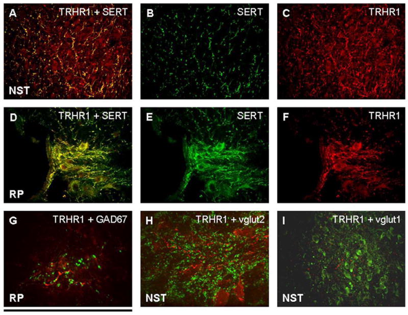
A, B, and C: Merged image (A) demonstrating that practically all SERT-ir fibers (B) in the NST possess TRHR1 receptors (C).
D, E, and F: Merged image (D) of staining in the raphe pallidus demonstrating that nearly all SERT-ir fibers and neurons (E) possess TRHR1 receptors (F).
G: GAD67-ir (GABA phenotype; green label) fibers in the RP are in the vicinity of TRHR1 receptors (red) but are not co-localized.
H: In the NST, vglut 2-ir fibers and varicosities (green) are in the vicinity of TRHR1 receptors (red) but are not co-localized.
I: Similarly, vglut 1-ir fibers and varicosities (green) in the NST are in the vicinity of TRHR1 receptors (red) but are not co-localized.
Scale bar: A–F, I = 150 microns; G, H = 20 microns.