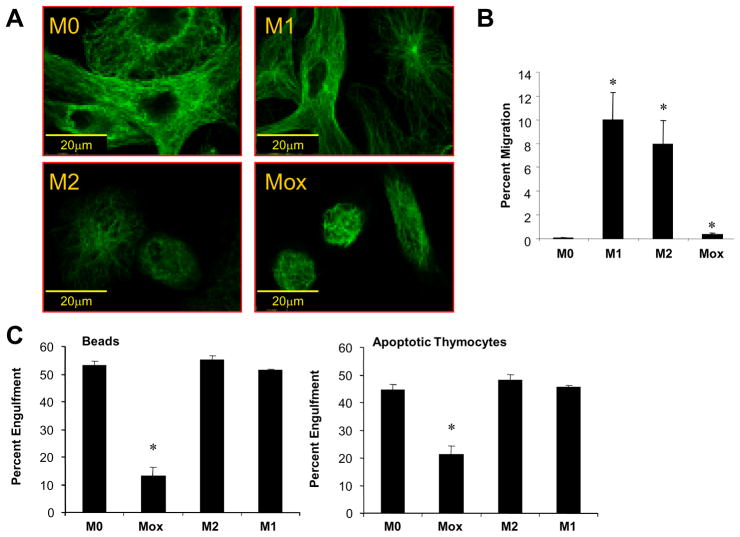
Figure 1. Mox macrophages represent a functionally distinct phenotype.
Bone marrow-derived macrophages were polarized with either 10U/ml IFNγ plus 1μg/ml LPS (M1) or 10ng/ml IL-4 (M2), 50μg/ml OxPAPC (Mox) or medium only (M0) for 18 hours. Cells were stained with an anti-tubulin antibody and analyzed by epifluorescence microscopy (A), bar indicates 20μm. (B) Migration of THP-1 cells to supernatants from macrophages treated as indicated, n=2. (C) Phagocytosis of carboxylate beads (left) and apoptotic murine thymocytes (right) by bone marrow-derived macrophages treated for 8 hrs as indicated, n=2.

