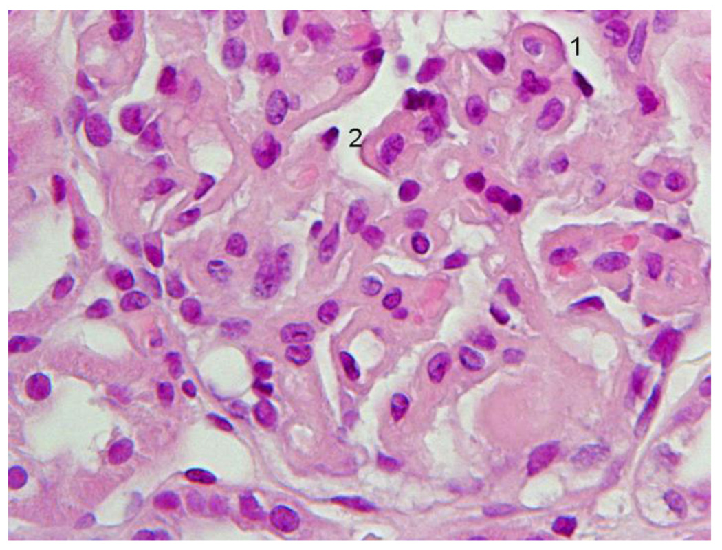
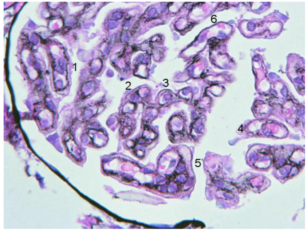
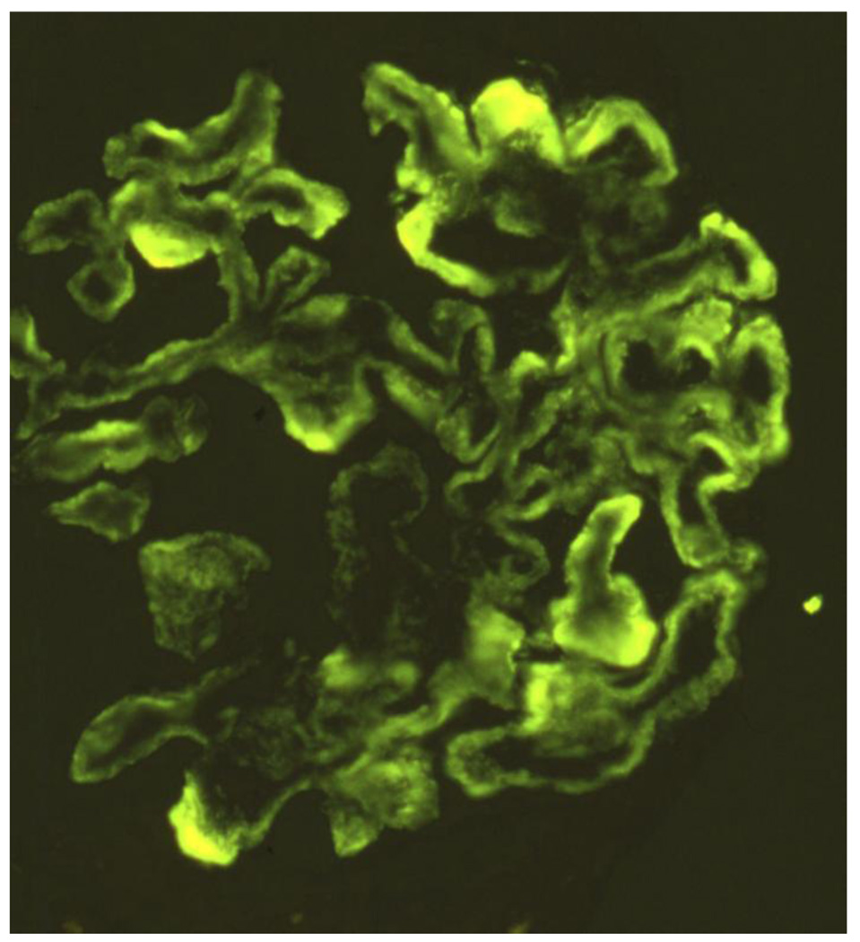
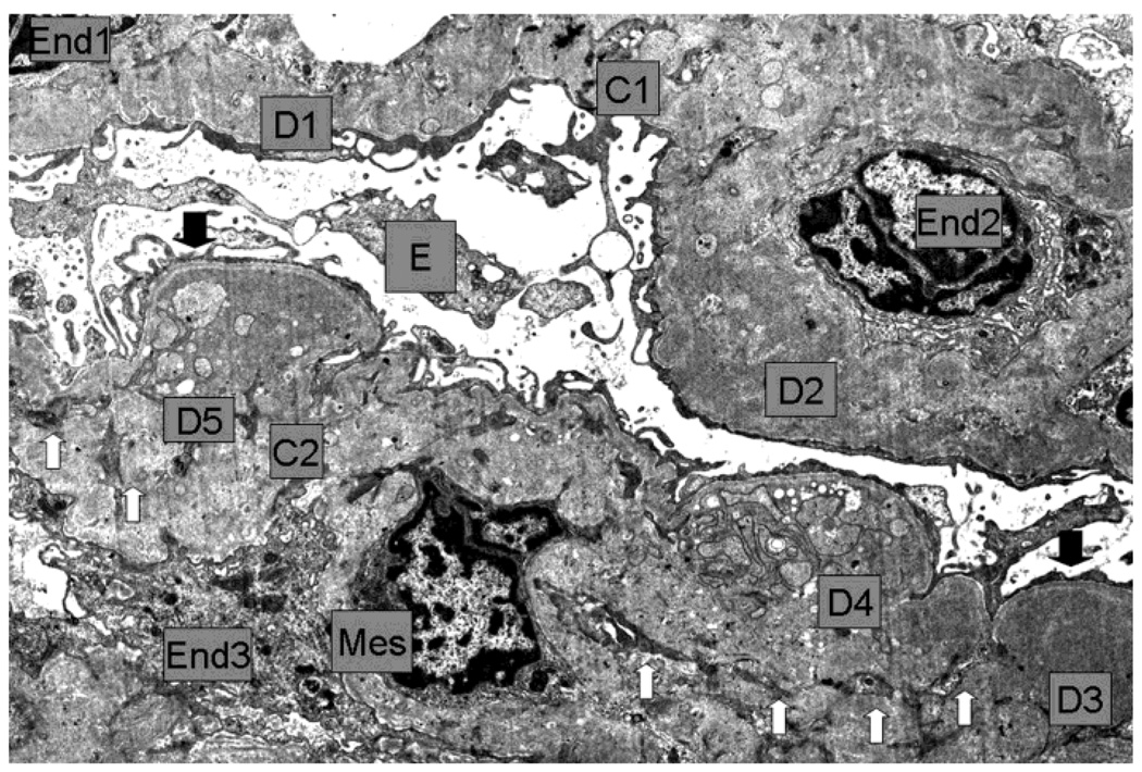
Figure 2.
First renal biopsy of the patient: Light microscopy (A) Silver stain (B), immunofluorescence (C), electromicroscopy (D)
2A Detail of the diffuse glomerulonephritis showing prominent generalized thickening of the capillary walls, sometimes with a broad glassy eosinophilic appearance (1, 2). There was slight to moderate increase in mesangial cells and expansion of the mesangial regions. Capillary lumina appear generally reduced. H+E.
2B. Within the thickened capillary walls the basement membrane appears irregular, interrupted or vacuolated (1, 3, 4). There are also several double contours some of which with a fairly thin outer contour (2, 3, 5). Any existing deposits within the wall appeared unstained with silver (5). One capillary wall has relatively normal wall (6). Mesangial matrix was mildly increased (5). Jones´s silver.
2C Intense confluent granular deposits of C4 with pseudolinear pattern globally distributed within the capillary walls and less intense mesangial staining. Large intense globular or semilunar collections in the wall were also seen.
2D Low magnification E.M: Capillaries (C1, C2) with endothelium (End1 to End3), Bowman´s space and visceral epithelium (E). In the thickened and deformed wall there are large, numerous moderately dense deposits (D1 to D5) more concentrated towards the subepithelial zone, but also extending through the whole wall (D2). Generally the dense deposits have uniform texture (D1 to D3) but some show complex irregular texture which includes less dense vesicular profiles, small clear spaces and groups of convoluted fine plasma membrane like structures (D4, D5). Generally there was no definition of the normal lamina densa of the basement membrane. Reactive type continuous thin lamina densa was seen along the epithelial side effectively separating the dense deposits from the podocytes (between black arrows). Clear cut formation of spikes was not seen. Reactive thin lamina densa on the endothelial side was ill defined and discontinuous. Mesangial cell (Mes) interposition was represented by multiple small cytoplasmic processes (white arrows) within the thickened capillary wall. There was severe thinning of podocytes and extensive effacement of foot processes. Negative magnification 2500×.


