Figure 4.
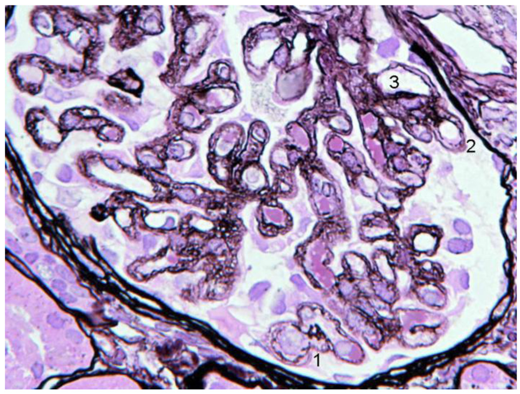
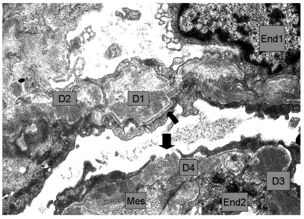
Second Biopsy: silver stain LM (A), EM (B)
4A. The basement membrane was generally irregular, sometimes interrupted (1,2) and double contours were present (3). The vacuolated or moth eaten profile of the walls, seen in the first biopsy, was widespread and also present in the paramesangium. Capillaries with normal single layer basement membrane were not seen. The glomerular capsule appeared marginally thicker than in the first biopsy.
4B. The walls of 2 capillaries with endothelial cells (End1 and End2) contained several dense deposits (examples D1 to D4). D1 appeared rarified and surrounded by thin reactive basement membrane which appeared well-defined and continuous in the subepithelium (thin arrow) and discontinuous in the subendothelium. D2 made direct contact with the podocyte. D3 had fairly uniform high density and was also partly surrounded by subepithelial basement membrane. There was a gap between podocyte processes leaving D4 in direct contact with Bowman´s space. Mesangial cell cytoplasm (Mes) interposition was seen in the wall near D4 and other deposits. Negative 6000×


