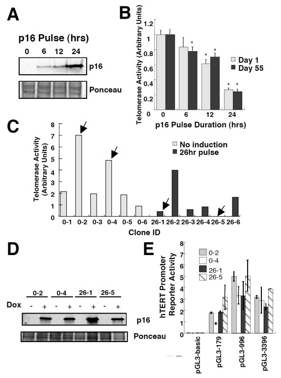
Fig. 4.
Time-dependent repression of telomerase activity following p16 induction is not due to loss or gain of diffusible transcriptional activators/repressors. (A) MCF7-tetR-tetp16 cells were treated with doxycycline for times indicated and harvested. p16 levels were determined by immunoblotting. (B) MCF7-tetR-tetp16 cells were treated with doxycycline for times indicated and grown under normal conditions. The cultures were divided when sub-confluent. At days 1 and 55 of the experiment, the cells were harvested and telomerase activity was determined. Mean values and standard deviations are shown (n=3). Values significantly different from the untreated control (p < 0.05, Student’s t-test) are denoted with a *. (C) MCF7-tetR-tetp16 cells were treated with doxycycline for 26 hours, seeded at clonal densities and expended into cell lines. Six doxycycline treated clones and 6 control clones were analyzed for telomerase activity. Arrows indicate telomerase activity in clones used in subsequent experiments. (D) p16 expression levels in MCF7-tetR-tetp16 clones were determined by immunoblotting. Negative control: parental MCF7; positive control: MDA-MB468. (E) Indicated cells were transfected with plasmids encoding luciferase under the control of hTERT promoter sequences of indicated lengths. hTERT promoter activity was measured and normalized to that of a co-transfected promoterless Renilla construct. Mean values and standard deviations are shown (n=3).

