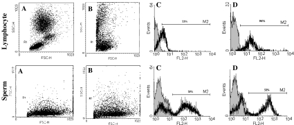
Fig. 2.
Lymphocytes and sperm treated with purified mouse anti human CD95 mAb followed by incubation with rabbit complement: a) Gate of lymphocyte and sperm before treatment, b) Gate of lymphocyte and sperm after treatment with CD59 mAb followed by incubation with rabbit complement, c) Histogram of PI in untreated lymphocytes and sperm and d) Histogram of PI lymphocytes and sperm after treatment with CD59 mAb followed by incubation with rabbit complement

