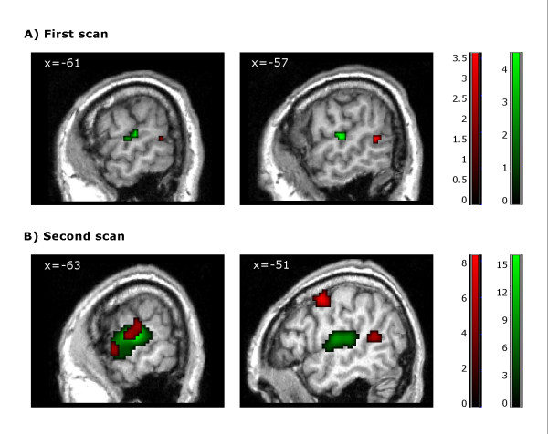
Figure 1.
Task -related activations. Patient's brain activation in the first (A) and second (B) scans for the contrast 'forward > backward narratives' (red) and the contrast 'narratives > silence' (green). Results are thresholded at p < 0.05 FDR-corrected and mapped on the patient's brain. Notice that the cluster displayed in the first scan for the contrast 'forward > backward narratives' is thresholded at p < 0.05 FDR-small volume correction. Color-bars indicate t statistic values. Numbers on the left superior corner of each image refer to the MNI-coordinate of the peak maxima in the x axis for each contrast.

