
Figure 2a:
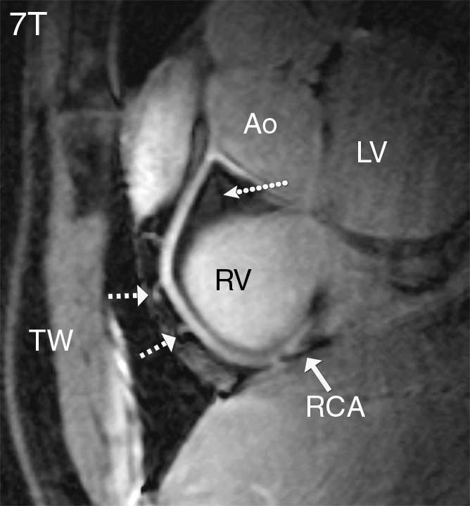
Coronary MR angiograms of the RCA obtained at (a) 7 T and (b) 3 T in the same healthy 18-year-old man (double oblique volume targeted plane parallel to the RCA). Improved suppression of the epicardial fat (long dotted arrow) with high contrast between the blood and epicardial fat is visible at 7 T. At both field strengths, a number of small branching vessels are depicted (short dashed arrows). Also at 7 T, a long portion of the RCA is visible (solid arrow = distal part of RCA). Ao = aortic root, LV = left ventricle, RV = right ventricle, TW = thoracic wall.