
Figure 1b:
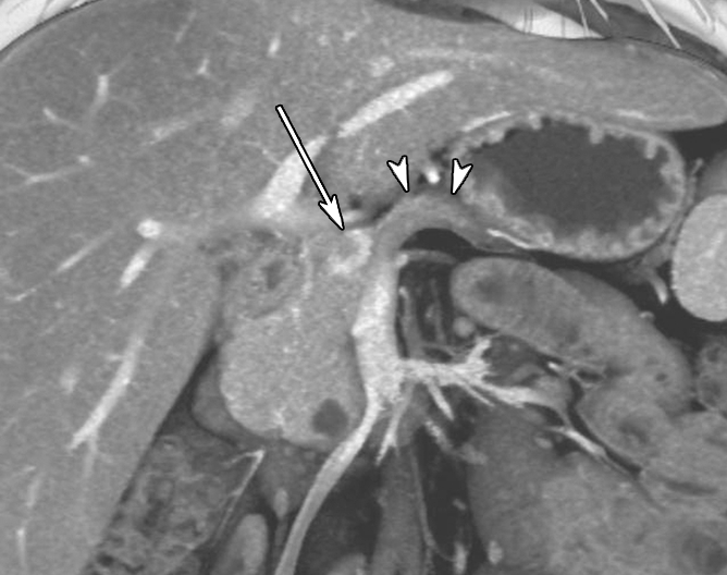
Contrast material–enhanced CT images in patient 1 (29-year-old woman). (a) Axial arterial phase image shows marked atrophy of the pancreatic body (arrowheads) (anteroposterior dimension = 5 mm). Arrow = small enhanced liver mass (metastasis). (b) Venous phase anterior volume-rendered image shows enhanced mass (arrow) with upstream pancreatic atrophy (arrowheads). A separate small cystic lesion (pathologic finding: oligocystic serous cystadenoma) is incidentally shown in the inferior aspect of the pancreatic head.