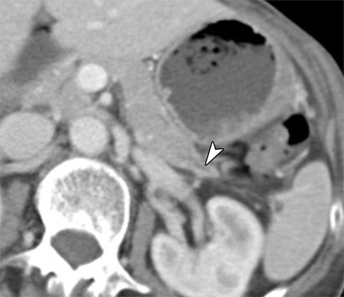
Figure 3b:

(a) Oblique axial arterial phase, (b) axial venous phase, and (c) oblique coronal CT images in patient 5 (64-year-old woman) show minimally dilated main pancreatic duct (arrowheads) in a pancreatic tail with an atrophic truncated appearance. No discrete mass is shown. There is a subtle hypoenhanced area (arrows) in the pancreatic tail.
