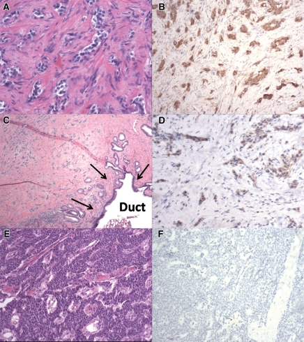
Figure 5:
Photomicrographs in patients 3 (A, B), 6 (C, D), and 2 (E, F). A, Sample shows well-differentiated endocrine neoplasm with marked stromal fibrosis. (Hematoxylin-eosin [H-E] stain.) B, D, Samples show strong serotonin immunoreactivity of neoplastic cells, with no labeling in stromal cells. C, Sample from downstream dilatation of the pancreatic duct shows well-differentiated endocrine neoplasm with marked stromal fibrosis extending into a main pancreatic duct (arrows). (H-E stain.) E, Sample shows a hypercellular well-differentiated endocrine neoplasm with minimal stromal fibrosis. (H-E stain.) F, Sample shows negative serotonin immunoreactivity of neoplastic cells. (Original magnification in A, ×200; in B, D, E, F, ×100; in C, ×20.)

