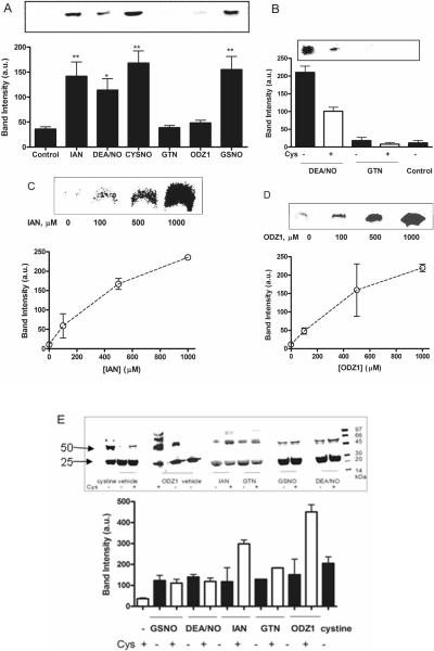
Fig. 2. Comparison of GST-P1 nitrosation and oxidation by NO-donors.
(A) BST analysis of GST-P1 (20 μM) treated with NO-donors (500 μM) at 37 °C for 30 min. ANOVA analysis with Dunnett's post test relative to vehicle control: ** p<0.001; * p<0.005. Response to IAN, GSNO, CysNO and DEA/NO was not significantly different. (B) BST analysis of GST-P1 (20 μM) untreated (control) or incubated with NO-donor (500 μM) in the presence and absence of Cys (2 mM). Quantitation was by image densitometry from blots. (C) BST analysis of GST-P1 (20 μM) nitrosation after incubation with varying concentrations of IAN at 37 °C for 30 min. (D) BST analysis of GST-P1 (20 μM) nitrosation after incubation with varying concentrations of ODZ1. (E) Non-reducing SDS-PAGE analysis of GST-P1 oxidation to dimer (and higher oligomers) by NO-donors (500 μM) visualized by Coomassie blue staining. GST-P1(20 μM) was incubated with NO-donors (500 μM) or cystine (1 mM) in the presence and absence of Cys (2 mM) at 37 °C for 30 min. Oxidation of GST-P1 was significantly increased in the presence of cysteine for IAN, GTN, and ODZ1, but not other NO-donors. Quantitation was by image densitometry from blots; upper panels show representative blots. Data show mean and s.d. in arbitrary units for triplicate experiments, which for oxidation/dimerization were normalized to the untreated control as 0%.

