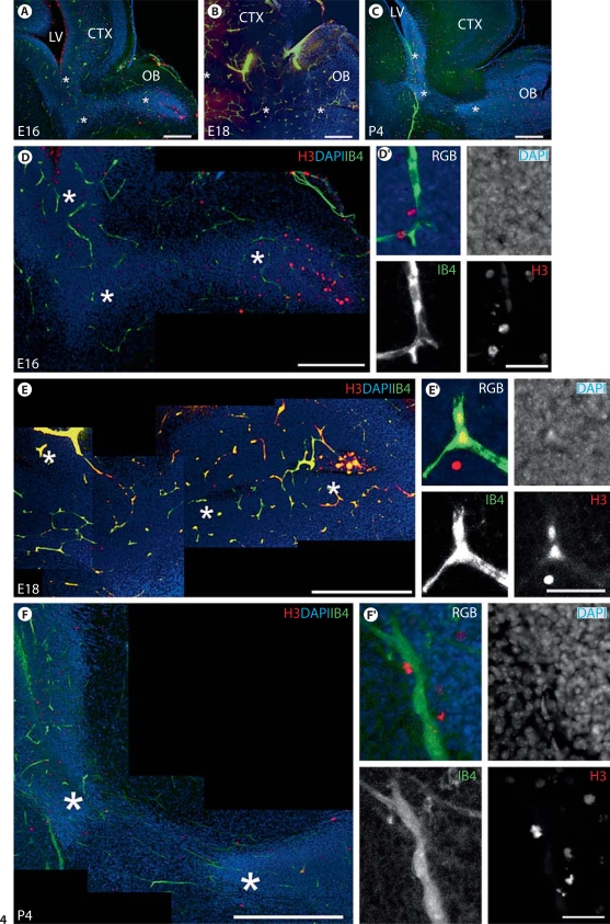
Fig. 4.
Proliferative cells in the RMS lie close to blood vessels at embryonic and early postnatal ages. A-C Low-power fluorescent photomicrographs of the rostral forebrain region at E16, E18 and P4 reveal the morphological development of the RMS in relation to other forebrain structures. A, B Scale bars = 200 μm. C Scale bar = 500 μm. CTX = Cortex; LV = lateral ventricle; OB = olfactory bulb. Asterisks mark the corresponding RMS at E16, E18 and P4 shown in D-F, respectively. D-F Confocal stacks (optical thickness 2.4 or 4.8 μm) of E16, E18 and P4 RMS, respectively. At E16 and E18, mitotic cells (H3+) are located in close association with blood vessels (IB4+) in the RMS despite vascular plexi at these ages showing no apparent organization with respect to the tangential course of the RMS (densely stained with DAPI). At P4, blood vessels align longitudinally along the direction of the RMS, and mitotic cells, as expected, are intimately associated with blood vessels. D Scale bar = 200 μm. E Scale bar = 300 μm. F Scale bar = 400 μm. D′-F′ High-magnification photomicrographs highlighting the close relationship between mitotic cells and blood vessels. Scale bars = 30 μm.

