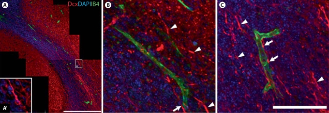
Fig. 6.
Relationship between migratory neuroblasts (Dcx+) and blood vessels (IB4+) in P4 RMS. A Montage of confocal images taken in a single optical plane gives an overview of the morphological structure of the RMS. Note that most postmitotic neurons outside the RMS retain Dcx immunofluorescence at this age. A′ Example of Dcx+ neuroblast shown at higher magnification. Scale bar = 300 μm. B, C High-power confocal photomicrographs show a few examples of Dcx+ neuroblasts intertwining with blood vessels in the RMS (arrows). However, there is no sign of an intimate association with blood vessels for the vast majority of Dcx+ neuroblasts (arrowheads) migrating in the P4 RMS. This therefore suggests that neuroblasts do not preferentially migrate along blood vessels. Scale bar = 100 μm.

