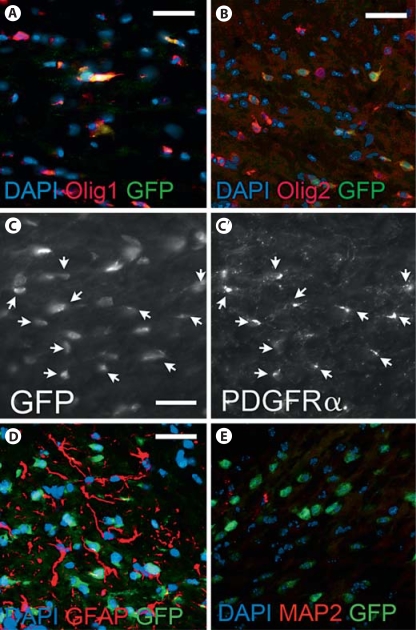
Fig. 3.
The Olig1-EGFP mouse reports Olig1+, Olig2+ cells and PDGFRα+ cells accurately. A Double immunofluorescence for Olig1+ cells and GFP+ cells at P14 shows all GFP+ cells co-labeling with Olig1 antibody within the corpus callosum [DAPI (blue), Olig1 (red), GFP (green), magnification ×40, scale bar = 50 μm]. However, not all Olig1+ cells were co-labeled by anti-GFP antibody. B Double immunofluorescence for Olig2+ cells and GFP at P14 shows many GFP+ cells co-labeling with Olig2 antibody within the corpus callosum [DAPI (blue), Olig2 (red), GFP (green), magnification ×40, scale bar = 50 μm]. C, C′ Double immunofluorescence for PDGFRα+ cells and GFP+ cells at P14 showed extensive overlap of cells within the corpus callosum [magnification ×40, scale bar = 50 μm, arrows indicate co-labeled cells]. GFP+ cells never co-labeled with antibody to GFAP at P16 [DAPI (blue), GFAP (red), GFP (green), magnification ×40, scale bar = 50 μm, n = 3] (D); nor with antibody to MAP2 at P14 [DAPI (blue), MAP2 (red), GFP (green), magnification ×40, scale bar = 50 μm, n = 3] (E).

