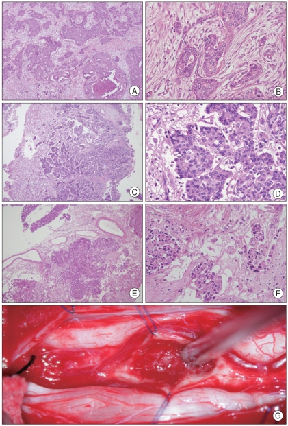
Fig. 3.
The primary tumor was infiltrating ductal carcinoma of breast. The lower power view (A : 40×) shows infiltrating carcinoma with ductal carcinoma in situ. High power view (B :×200) shows infiltrating carcinoma with focal tubular patterns. The histological features of the cervical ISCM and conus medullaris ISCM second metastasis to the spinal cord are identical to the primary tumor. C (×40) and D (×200) : Cervical ISCM. E (×40) and F (×200) : Conus medullaris ISCM. G shows intraoperative finding of conus medullaris ISCM.

