Figure 1.
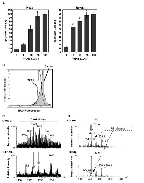
TRAIL induces changes in mitochondrial cardiolipin. A, TRAIL-induced apoptosis: HeLa and Jurkat cells were treated with TRAIL at various concentrations for 24 hours. Cells were collected and the extent of apoptosis was determined by flow cytometry of the sub-G1 population. Columns, mean of three independent determinations; bars, SD. *, P < 0.05, statistically significant two-sided Wilcoxon test compared with control untreated cells. B, TRAIL-induced changes in cardiolipin: Jurkat cells were incubated with TRAIL (10 ng/mL) for 1 hour and then stained with NAO, the fluorescence of which was analyzed by flow cytometry. C, electrospray MS profile of mitochondrial lipid extracts: untreated (top) and TRAIL-treated Jurkat cells (10 ng/mL for 1 hour; bottom). Although the reference intensity of the dominant phosphatidylcholine species was comparatively higher for the TRAIL-treated sample than the control sample, the MS profile of the cardiolipin region was recorded to an equivalent signal-to-noise ratio to emphasize both quantitative and qualitative changes in cardiolipin species. D, electrospray MS spectra of the major mitochondrial phospholipids dominated by phosphatidylcholine (PC) species: The spectra were normalized to the intensity of the dominant palmitoyl,arachidonyl-phosphatidylcholine at 782 m/z, although this lipid showed some TRAIL-induced increase with respect to other arachidonyl-containing species (e.g., that at 808 m/z) and internal references like the major phosphatidylethanolamine (PE) species at 768 m/z. Note the TRAIL-induced severe depletion of mitochondrial palmitoyl,oleoyl-phosphatidylcholine at 760 m/z (thick arrows).

