Figure 4.
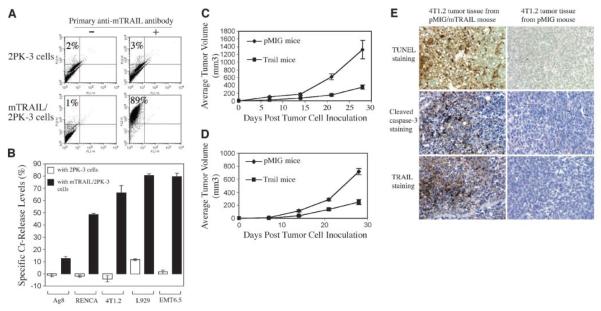
A, mTRAIL protein expression on mTRAIL/2PK-3 cells. 2PK-3 cells and mTRAIL/2PK-3 cells were incubated with either nonimmune rat IgG (−) or N2B2 anti-mTRAIL (+) antibody followed by biotin-conjugated goat anti-rat Ig and phycoerythrin conjugate and analyzed by FACS channel 2 for phycoerythrin. mTRAIL/2PK-3 cells were used to select mTRAIL-sensitive mouse tumor cells. B, Cr release assay for selection of mTRAIL-sensitive mouse tumor cell lines. Columns, result from four replicate samples (n = 4). P < 0.01, for samples with 2PK-3 cells versus samples with mTRAIL/2PK-3 cells (Student's t test). Four experiments were done with consistently similar results. C, EMT6.5 tumor growth in bone marrow recipient mice. Points, mean from 9 mice in pMIG group (n = 9) and from 12 mice in pMIG/mTRAIL group (TRAIL mice; n = 12). P < 0.005, by day 28 (Student's t test). Experiment repeated thrice with similar results. D, 4T1.2 tumor growth in bone marrow recipient mice. Points, mean from 12 mice in each group (n = 12; labeled as the same in Fig. 4C). P < 0.005, by day 28 (Student's t test). E, inhibition of tumor growth in mTRAIL-transduced mice involves mTRAIL-induced apoptosis: evidence by immunohistochemistry staining. The tumor tissues were harvested by the 4th week after the tumor cell inoculation and immediately processed in TUNEL staining, cleaved caspase-3 staining, and mTRAIL staining. The tumor tissue from pMIG/mTRAIL-transduced mice shows intensive apoptosis by TUNEL staining, highly positive staining of cleaved caspase-3, and presence of high level of mTRAIL protein. Those positive staining results cannot be observed in the tumor tissues from pMIG-transduced mice.

