Fig. 2.

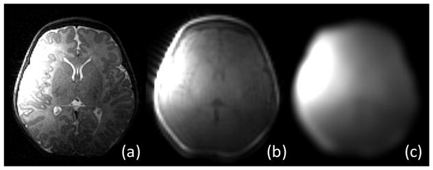
Illustrations of (a) coil image, (b) coil profile image, and (c) estimated sensitivity map. Note that, to reach (c), non-brain tissues are first removed to better concentrate on the brain parenchyma.

Illustrations of (a) coil image, (b) coil profile image, and (c) estimated sensitivity map. Note that, to reach (c), non-brain tissues are first removed to better concentrate on the brain parenchyma.