
Fig. 2. The misfolded mutant SOD1 specifically co-precipitates with VDAC1 in spinal cord mitochondria.
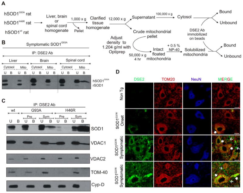
(A) Schematic showing the isolation of cytosolic and mitochondrial fractions. (B) Liver, brain and spinal cord cytosolic and mitochondrial fractions were purified from symptomatic rats expressing hSOD1G93A and the fractions were subjected to immunoprecipitation using DSE2 (3H1), a monoclonal antibody only recognizing misfolded SOD1 (Vande Velde et al., 2008). The immunoprecipitates were immunoblotted using an SOD1 antibody. (C) Isolated floated mitochondria from hSOD1wt, hSOD1G93A and hSOD1H46R rat spinal cords (from pre-symptomatic and symptomatic animals) were immunoprecipitated with DSE2 (3H1), and the immunoprecipitates were immunoblotted using VDAC1, VDAC2, TOM-40 and cyclophilin-D antibodies. SOD1 immunoprecipitation was confirmed by reprobing the membrane with an SOD1 antibody (top). (D) Immunohistochemical detection of misfolded SOD1 using DSE2 antibody shows that misfolded SOD1 (green) colocalizes with TOM20 (red), a mitochondrial outer membrane protein in a subset of spinal cord neurons assessed using NeuN (blue), a neuronal marker as highlighted by filled arrows. DSE2 positive staining can be detected in some neurons at onset and significantly increases with the appearance of disease symptoms.
Of note DSE2 staining is not restricted to neuronal mitochondria but is also detected in non-neuronal cells and the extracellular space as shown with thin arrows. No DSE2 staining was detected in neurons of 1 year old non transgenic control mice (Non Tg). Scale bar: 10 μm. Abbreviation: U, unbound fraction (20 %); B, bound fraction; Pre, pre-symptomatic; Sym, symptomatic.