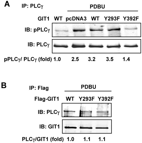
Figure 5. Role of GIT1 tyrosine phosphorylation in PLCγ activation and GIT1-PLCγ interaction.
(A) Effects of GIT1 tyrosine mutants on PLCγ activation. HEK293 cells were co-transfected with PLCγ together with pCDNA3 vector, Flag-GIT1 (WT), Flag-GIT1 (Y293F), or Flag-GIT1 (Y392F) (B) for 24 hours and then starved for 6 hours. Cells were treated with or without 1μM PDBU for 10 min and cell lysates were immunoprecipitated with PLCγ antibody and immunoblotted with pPLCγ antibody (top panel). To confirm equal protein immunoprecipitation, the blot was reprobed with PLCγ antibody (lower panel). (B) Effects of GIT1 tyrosine mutants on GIT1-PLCγ interaction. HEK293 cells were co-transfected with PLCγ together with Flag-GIT1 (WT), Flag-GIT1 (Y293F), or Flag-GIT1 (Y392F) for 24 hours and starved for 6 hours. After treatment with 1μM PDBU for 10 min, cell lysates were immunoprecipitated with Flag antibody for GIT1 and then immunoblotted with PLCγ antibody. To confirm equal protein immunoprecipitation, the blot was reprobed with GIT1 antibody. The blots were analyzed by densitometry using LiCor software. Fold changes normalized to the first lane are shown below the blots (n=2-3).

