
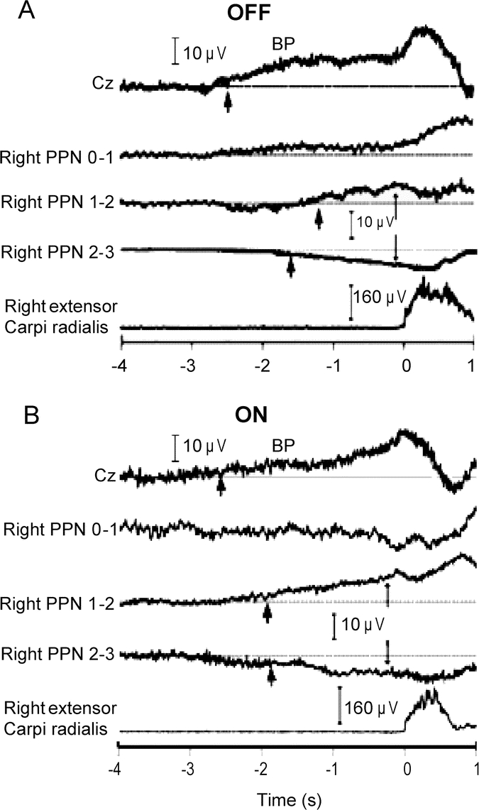
Figure 1 Examples for scalp and bipolar PPNR MRP recordings
Recordings from self-paced ipsilateral wrist extension movements from patient 1 in OFF (A) and ON (B) states. Cz is the vertex scalp electrode, and 0–1, 1–2, and 2–3 represent bipolar montage of the quadripolar deep brain stimulation (DBS) electrodes at the pedunculopontine nucleus region (PPNR). The lowest traces show the rectified and averaged EMG activity of the right extensor carpi radialis muscle (A and B). The horizontal lines represent the baselines. The scalp recording shows a slow, negative movement-related potential (MRP) (Bereitschaftspotential [BP]). The thick arrows point to MRP onsets. The thin arrows signify phase reversals between adjacent DBS contact pairs. The BP onset latencies (approximately −2.5 seconds) for the scalp electrodes were similar between OFF and ON medication states. For the PPNR electrodes, the BP onset occurred at approximately −2 seconds in the OFF and ON states. PPN = pedunculopontine nucleus.