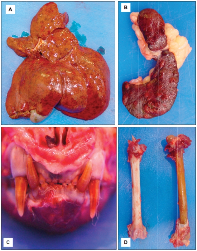
Figure 1.
A — The patient’s liver showing numerous areas of red discoloration. B — The spleen with numerous white nodules throughout the parenchyma. C — The patient’s skull showing brown discoloration of the dentition. D — Brown discoloration of the affected patient’s femur (right) compared to a femur from a cat that was not affected with porphyria (left).

