
Figure 1.
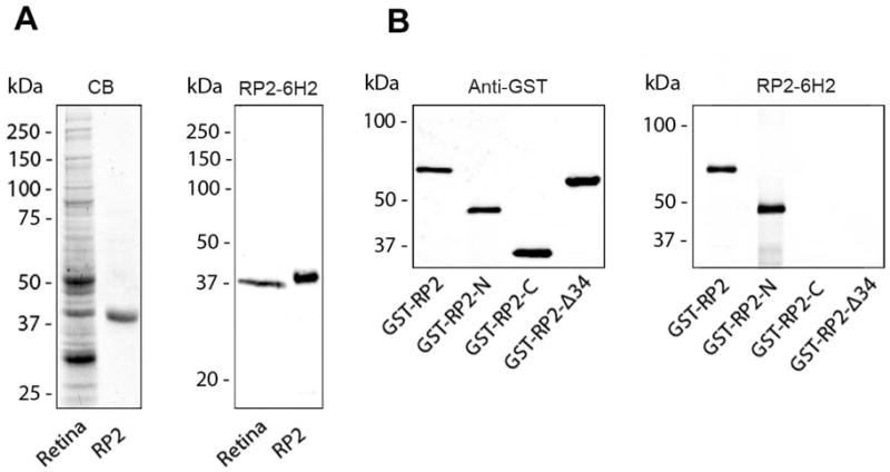
Characterization of RP2-6H2 antibody using bovine retinal extract and His-tagged human RP2. (A) Coomassie blue-stained gel (CB) and a western blot of a retinal extract (retina) and purified His-tagged RP2 (RP2) labeled with the RP2-6H2 antibody demonstrating the specificity of the antibody. (B) A series of GST-RP2 fusion proteins were used to map the epitope of RP2-6H2 antibody. The RP2-6H2 antibody labels the full length (GST-RP2) and the N-terminal fusion protein (GST-RP2-N) but not the C-terminal fusion protein (GST-RP2-C) or GST-RP2 lacking the first 34 amino acids (GST-RP2-Δ34) as shown in the right panel. The GST-fusion proteins were identified in the western blot labeled with an anti-GST antibody.