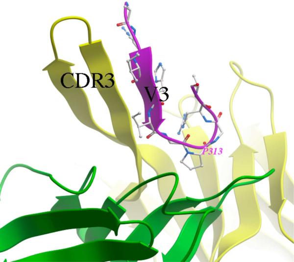
Figure 1.
The V3 peptide fragment (shown in magenta ribbon representation and sticks) bound to the broadly neutralizing mAb 447-52D (light and heavy chains are shown as green and yellow ribbons, respectively). Formation of a three-strand beta-sheet composed of two strands of the heavy chain CDR3 hairpin and one V3 strand, as well as tight binding of the conserved residue P313 in the GPGR motif at the tip of the loop can be observed. All molecular graphics images were prepared in Molsoft ICM-Pro.

