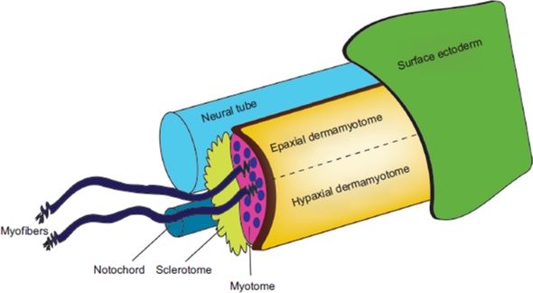
Figure 2.
Caricature showing the structures in the skeletal muscle. In general, the main skeletal muscle anatomy consists of the dermomyotome, myotome and sclerotome, and is conserved throughout species. The dermomyotome is the source of the primary myotome, as well as contributing to the formation of the dermis, the endothelial and smooth muscle cells. The dermomyotome is divided into epaxial and hypaxial domains, which give rise to the epaxial muscle (deep muscle of the back) and hypaxial muscles (appendicular musculature, abdominal muscles, diaphragm, hypoglossal chords) respectively.

