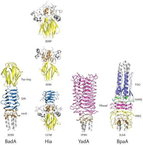
Figure 5. Comparison of architecture of TAA head domain sequence motifs.
Trimeric structures are shown for the TAA head domains of BadA from Bartonella henselae (PDB ID 3D9X [11]), HiaBD2 (PDB ID 3EMF [9]), KG1-W3 (PDB ID 3EMI [9]), and HiaBD1 (PDB ID 1S7M [10]), from Haemophilus influenzae, YadA from Yersinia enterocolitica (PDB ID 1P9H [7]), and BpaA from B. pseudomallei (PDB ID 3LAA). Trp-ring motifs are shown in yellow, GIN motifs are shown in light blue, neck regions are shown in brown, left handed β-roll Ylhead repeats are shown in magenta, FGG motifs are shown in dark blue, HANS motifs are shown in green, HIN2 motifs are shown in orange, and other regions in gray.

