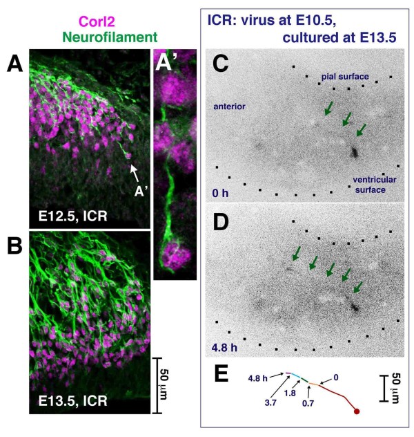
Figure 5.
Early axonogenesis in nascent Purkinje cells. (A, B) Anti-Neurofilament and anti-Corl2 double immunostaining showing that many Purkinje cells have axon-like fibers running either radially or tangentially in E12.5 and E13.5 cerebella. (C-E) Time-lapse observation of an E10.5-born Purkinje-like cell extending an axon-like fiber (arrows) to the anterior side in an E13.5 cerebellar slice (see also Additional files 4B and 5). Although not immunostained, it is highly likely that the cell in this time-lapse is a Purkinje cell based on our in vivo data, which indicate that most GFP+ cells within a deep cerebellar region sandwiched by the outermost territory for DN neurons and the VZ are Lhx1/5+ (96% in the E10.5 to E13.5 analysis; n = 45/47).

