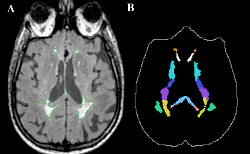
Figure 1.

Example illustrating some of the white matter ROIs analyzed in the study. Panel A shows a grayscale FLAIR image with the WMH outlined in green. Panel B shows the ROIs mask corresponding to panel A that we used for determining the regional WMH burden. In panel B the intracranial cavity mask is outlined in white. Solid colors in panel B (top to bottom): orange (anterior corona radiata, ACR), white (genu of the corpus callosum, GCC), green-blue (anterior limb of the internal capsule, ALIC), dark blue (posterior limb of the internal capsule, PLIC), purple (superior corona radiata, SCR), yellow (posterior corona radiata, PCR), light blue (body of the corpus callosum, BCC), pink (splenium of the corpus callosum, SCC), green (superior longitudinal fasciculus, SLF).
