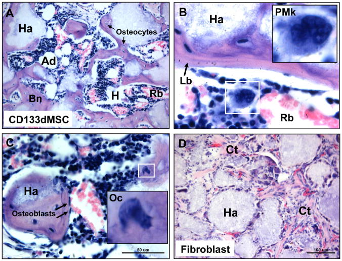
Fig. 3. Histology of cell-seeded constructs 60 days after implantation.
A) H & E stain of CD133BMSC-seeded construct showing HME formation with blood forming hematopoietic cells (Hp), enucleated red blood cells (Rb), and adipocytes (Ad), that are interspersed throughout bone (Bn) (10X magnification). Note that bone surrounds the HA/TCP carrier (Ha). Osteocytes (Os) were observed residing within lacunae (arrows). B) Concentrically-layered lamellar bone (Lb, arrow) had formed on HA/TCP surfaces (Ha) (40X magnification). Inset: promegakaryocyte (PMk). C) Erythropoiesis at various stages was observed throughout the HMEs. Inset: osteoclast (Oc). Arrows: osteoblasts. D) HA/TCP/fibrin construct seeded with 2 × 10⁶ human fibroblasts. Fibroblasts adhered to the HA/TCP matrix (Ha) formed connective tissue (Ct) containing microvasculature throughout the implant, but did not support hematopoiesis.

