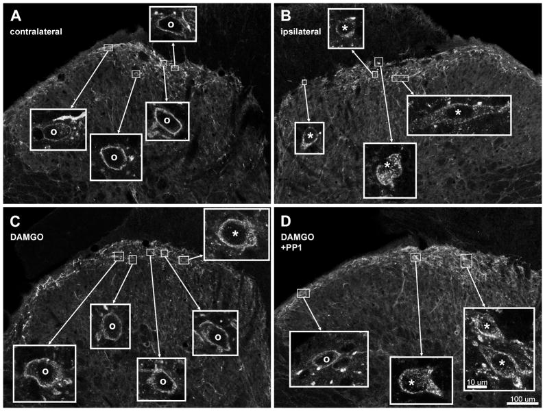
Fig. 1. Confocal images of NK1R neurons in lamina I after noxious stimulation of the hind paw in vivo.
Rats received a noxious stimulus (clamp for 30 sec) in the hind paw to induce substance P release and were fixed 10 min later for NK1R immunohistochemistry. Images were taken from sections of the L4–L5 spinal segments. A, B: Rats received an intrathecal injection of saline 10 min before the stimulus; A and B correspond to the contralateral and ipsilateral dorsal horns, respectively, of the same histological section. B: Rats received an intrathecal injection of DAMGO (2 nmol) 10 min before the stimulus; ipsilateral dorsal horn. C: Rats received intrathecal injections of DAMGO (2 nmol) and PP1 (10 nmol) 10 min before the stimulus and of PP1 (10 nmol) 70 min before the stimulus; ipsilateral dorsal horn. Main panels: images taken with a 10x objective, with a voxel size of 830 × 830 × 5983 nm and 2 confocal planes. Insets: images of lamina I neurons taken with a 63x objective, with a voxel size of 132 × 132 × 383 nm and 3 confocal planes. Neurons with NK1R internalization are indicated with “*” and neurons without internalization by “o”. Scale bars (in panel D) are 100 μm for the main panels and 10 μm for the insets.

