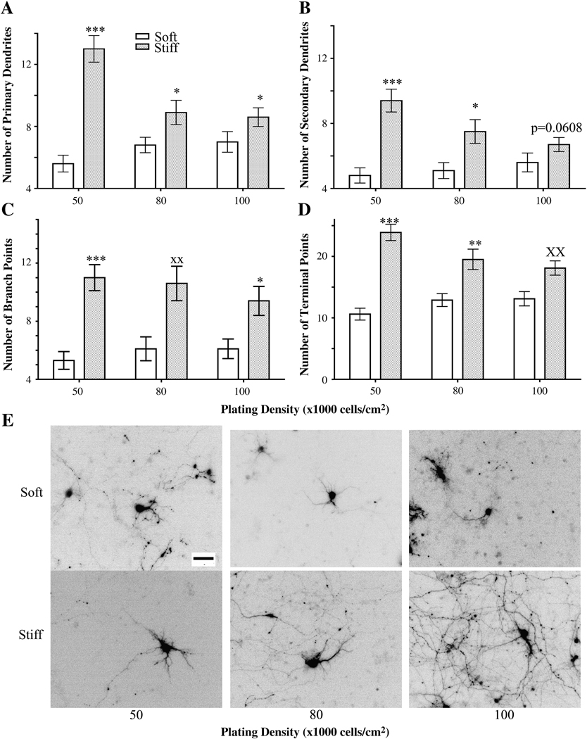
Figure 4.
Dendrite branching patterns of neurons plated at different initial densities and on different substrate rigidities as assessed on 12 DIV. Neurons plated on stiff gels had more primary (A) and secondary (B) dendrites than those plated on soft gels at the same initial cell density. ***p<0.0001 and *p<0.05 determined by Mann-Whitney Test. (C) Numbers of dendrite branch points increased in neurons plated on stiff gels compared to those plated on soft gels when plated at an initial density of 50,000 cells/cm2. ***p<0.0001 and *p<0.05 determined by Mann-Whitney Test. **p<0.01 determined by Student’s t-test. (D) Numbers of dendrite terminal points increased in neurons plated on stiff gels compared to those grown on soft gels when plated at an initial density of 50,000 cells/cm2. ***p<0.0001 and **p<0.01 determined by Mann-Whitney Test and xxp<0.01 determined by Student’s t-Test. (E) Representative, inverted images of GFP-expressing neurons quantitated in panels A–E. Data were averaged across four experiments. All data are represented as mean ± SEM. Numbers of cells for each condition are listed for 50,000, 80,000, and 100,000 cells/cm2, respectively. Soft gels: n= 28; 37; 34. Stiff gels: 35; 30; 33. Scale bar, 50 µm.

