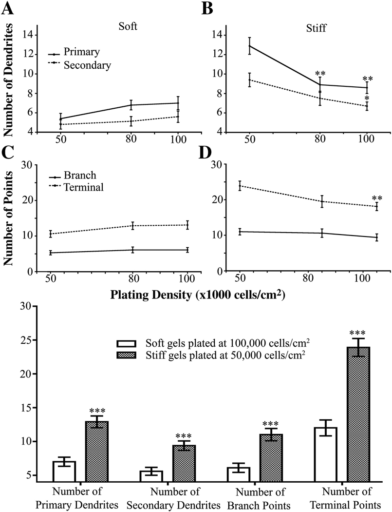
Figure 5.
The number of dendrites is independent of initial plating density for neurons grown on soft gels. (A) Neurons plated on soft gels showed no change in number of primary or secondary dendrites. (B) Neurons plated on stiff gels have less primary and secondary dendrites when plated at higher initial cell densities. **p<0.01 for primary dendrite number determined by ANOVA followed by Dunnett’s Multiple Comparison Test compared to initial plating density of 50,000 cells/cm2. *p<0.05 for secondary dendrites determined by Kruskal-Wallis Test followed by Dunn’s Multiple Comparison Test compared to initial plating density of 50,000 cells/cm2. (C) Number of dendrite branch and terminal points did not change in neurons plated at increasing cell densities on soft gels. (D) Number of dendrite branch points did not change, but number of terminal points decreased, in neurons plated at increasing cell densities on stiff gels. **p<0.01 determined by ANOVA followed by Dunnett’s Multiple Comparison Test compared to 50,000 cells/cm2. Data were averaged across four experiments All data are represented as mean ± SEM. Numbers of cells for each condition are listed for 50,000, 80,000, and 100,000 cells/cm2, respectively. Soft gels: n= 28; 37; 34. Stiff gels: 35; 30; 33.

